Diagnosis of Ringworm (Tinea Infections)
Ringworm is a common fungal infection of the skin, caused by dermatophytes. Despite its name, it has nothing to do with worms; the term comes from the ring-shaped rash it typically causes. Diagnosing ringworm is important to ensure proper treatment and to prevent the spread of the infection. Here’s how ringworm is typically diagnosed:
1. Clinical Examination
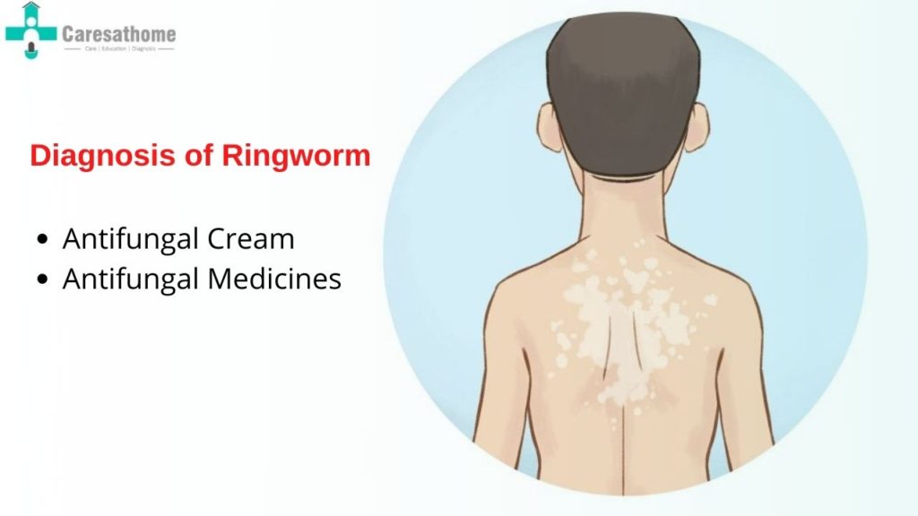
A healthcare provider will begin by examining the affected skin area. Ringworm often presents with characteristic features, including:
- Red, circular or ring-shaped rash with a clear center.
- Scaly, itchy patches on the skin.
- Raised edges that can appear inflamed.
- On the scalp, bald patches with scaling may appear, and in nails, thickening or discoloration can occur.
The appearance of the rash can be enough to make a preliminary diagnosis of ringworm, but additional tests may be used for confirmation.
2. Wood’s Lamp Test
A Wood’s lamp (ultraviolet light) may be used to examine the affected area. Certain types of ringworm, particularly those caused by Microsporum fungi, will fluoresce under the UV light, glowing a bright greenish-yellow. However, not all fungal species that cause ringworm will glow under a Wood’s lamp, so this test isn’t always conclusive.
3. Microscopic Examination (KOH Test)
For a more definitive diagnosis, a potassium hydroxide (KOH) test can be performed:
- A small sample of skin, hair, or nail from the affected area is collected by gently scraping the lesion.
- The sample is then treated with potassium hydroxide (KOH) and examined under a microscope.
- The KOH dissolves non-fungal cells, making the fungal elements, such as hyphae (branching filaments), easier to see.
This is a quick and reliable test to confirm the presence of dermatophytes.
4. Fungal Culture
In cases where the diagnosis is uncertain, or if the infection is not responding to treatment, a fungal culture may be done:
- Skin scrapings or other samples are placed in a growth medium and monitored for fungal growth over a period of days to weeks.
- The type of fungus can then be identified based on the growth and appearance of the culture.
Though this test is more time-consuming, it can be useful in cases of recurrent or severe infections to identify the exact fungal species causing the ringworm.
5. Biopsy and Histopathology
In rare or severe cases, a skin biopsy may be performed, especially if the diagnosis remains unclear or if the infection is not improving with treatment. A small piece of the affected skin is removed and examined under a microscope to check for fungal elements and tissue changes.
6. Dermatophyte Test Medium (DTM)
A Dermatophyte Test Medium is a type of culture used in some clinics for faster identification:
- The sample is placed on a special medium that changes color if dermatophytes are present.
- It provides quicker results than standard fungal cultures, although confirmation of the fungal species may still be needed.
Differential Diagnosis:
It’s important to differentiate ringworm from other skin conditions that may have similar symptoms, such as:
- Eczema (atopic dermatitis)
- Psoriasis
- Contact dermatitis
- Seborrheic dermatitis
- Pityriasis rosea
Proper diagnostic tests help avoid misdiagnosis and ensure appropriate antifungal treatment is administered.
Conclusion
Ringworm is typically diagnosed through a combination of clinical examination, KOH test, Wood’s lamp examination, and, if necessary, fungal culture or biopsy. Early and accurate diagnosis is key to preventing the spread of infection and achieving effective treatment.